
Select Dental Implants · Menifee, CA
Implant Technology at Select
Cone-beam 3D imaging, digital impressions, and CAD/CAM restorations — precision equipment used on every case, not just the brochure.
360°
full 3D view of tooth, bone, nerves and sinus
Minutes
digital impressions instead of trays
Every case
3D-planned before surgery
Cone-Beam 3D Scanner (CBCT)
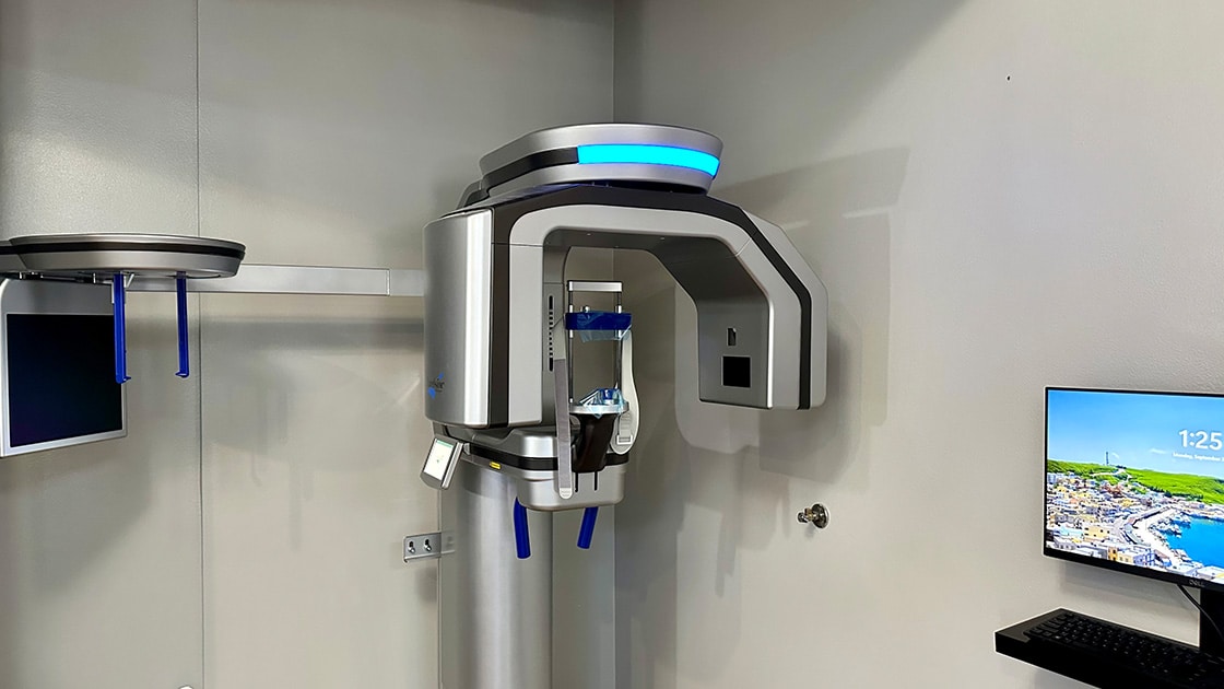
A traditional X-ray is only two-dimensional. Our cone-beam scanner gives the doctor a full 360-degree, 3D tomographic view of the tooth and all surrounding structures — bone, nerves, and sinuses — while using less radiation than traditional medical CT technology. The scan is fast, simple, and painless.
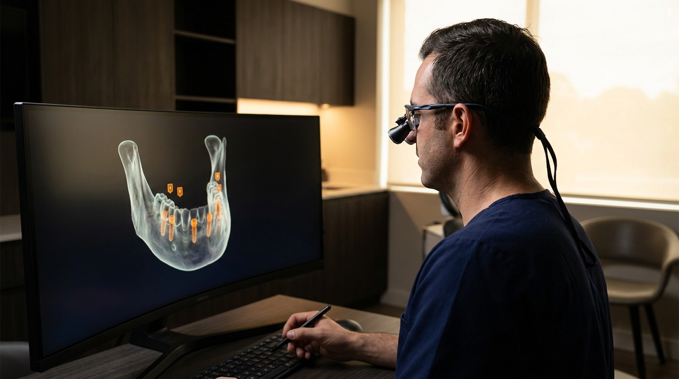
For implant patients this is the difference between estimating and knowing: bone volume measured to the millimeter, the inferior alveolar nerve mapped, sinus position confirmed, and implant placement planned digitally before surgery. Every implant case at Select starts here — that’s our actual scanner in the photo.
- Bone height, width and density measured precisely
- Nerve and sinus position mapped before any incision
- Implant size, angle and depth chosen digitally
- The basis for a printed surgical guide on guided cases
Digital Impressions & CAD/CAM Restorations
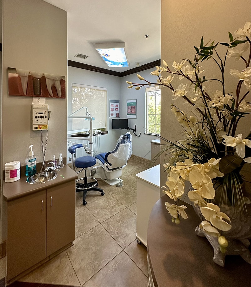
Impressions traditionally meant trays of soft, messy, often bad-tasting material hardening around your teeth. Our digital intraoral scanner replaces that with a comfortable, highly precise digital capture — the foundation for planning treatment and crafting final restorations.
CAD/CAM (computer-aided design and manufacturing) then fabricates precise inlays, onlays, crowns, and bridges: a digital impression feeds the design software, and the restoration is milled from a solid block of ceramic — durable, aesthetic, and well-fitted in fewer visits.
There’s a quieter benefit too. Your scan is a permanent digital record. If a crown is ever damaged years later, the file already exists — remaking it doesn’t start from scratch.
Guided Placement, and Why Millimeters Matter
Experienced surgeons place excellent implants freehand every day. What digital guidance adds is repeatability at the margins: sites close to nerves and sinuses, narrow ridges, multiple implants that must align to carry one bridge, and full-arch cases where four posts support every tooth.
Accurate placement isn’t cosmetic detail — spacing determines whether gum tissue forms naturally between teeth, whether the site can be cleaned, and whether bone survives long term. More on that in our guide to the 3-2 spacing rule.
Questions Patients Ask Us
Is a cone-beam scan safe? How much radiation?
A cone-beam CT uses substantially less radiation than a traditional medical CT scan of the same area, and the scan takes seconds. We take one when it changes the treatment decision — which for implant planning it essentially always does — and not as a routine add-on.
Do I still need messy impression trays?
For most procedures, no. A digital intraoral scanner captures your teeth with a small wand in a few minutes. Patients with strong gag reflexes in particular tend to find it a dramatic improvement over trays.
What does ‘guided surgery’ actually change for me?
Your implant position is decided on screen before surgery day and transferred to your mouth with a custom-printed guide. In practice that means a smaller opening, less time in the chair, and a position chosen for the final crown rather than improvised during surgery.
Can I see my own scan?
Yes, and we encourage it. Seeing your own jaw in three dimensions is usually the moment the treatment plan stops being abstract — bone volume becomes a number you can look at rather than a claim you have to take on faith.

See the Technology in Person
Ask for a look at your own 3D scan at your consultation — patients love it. Call (951) 672-6788.